Monday, 2 December 2024

How to Deal With Morbidly Adherent Placenta (MAP)

Managing a morbidly adherent placenta (MAP), which includes conditions like placenta accreta, increta, and percreta, is challenging and requires a multidisciplinary approach. Below are the key steps in the management process:


1. Antenatal Diagnosis 

Suspect MAP in all patients with previous cesarean or uterine surgery and if the placenta is anteriorly located. 

• Ultrasound with Doppler is the primary tool for diagnosing MAP.

• MRI may be used in uncertain cases or to assess the extent of invasion.

• Early diagnosis allows for better planning, including scheduling delivery in a tertiary care center.


2. Multidisciplinary Planning

Involves a team of:

• Obstetricians (Maternal-Fetal Medicine specialists if available)

• Anesthesiologists

• Interventional radiologists

• Neonatologists

• Urologists or general surgeons (if bladder or bowel involvement is suspected)

• Blood bank team

Planning includes:

• Counseling the patient about risks, delivery timing, and possible hysterectomy.

• Ensuring adequate blood products are available.


3. Timing and Mode of Delivery

• Elective cesarean section is typically scheduled around 34-36 weeks, after steroid administration for fetal lung maturity.

• Vaginal delivery is contraindicated due to the risk of severe hemorrhage.


4. Intraoperative Management

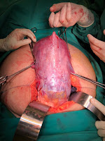
• Cesarean hysterectomy (CH) is the definitive treatment in most cases: Subtotal hysterectomy with minimise bleeding & injuries to the bladder in my opinion . Based on personal experience. 


• Deliver the baby via a classical uterine incision (avoiding the placenta).

• Leave the placenta in situ to minimize hemorrhage, before proceeding with CH

• Proceed with a planned Cesarean hysterectomy on step wise manner. 

• Uterine artery embolization or balloon occlusion catheters can be used to reduce blood loss prior to CH 

• Massive hemorrhage protocol should be in place, with cross-matched blood, platelets, and coagulation factors ready at the time of CH 


If hysterectomy is not performed:

• Consider leaving the placenta in situ with close follow-up, but this is reserved for selected cases and carries risks like infection or delayed hemorrhage.


5. Postoperative Care: HDU/  ICU 

• Monitor for complications such as:

• Infection

• Secondary hemorrhage

• Coagulopathy

• Administer thromboprophylaxis as per risk assessment.


Key Challenges

• Massive hemorrhage is the leading cause of maternal morbidity and mortality in MAP.

• Bladder injury is common in placenta percreta cases.

• Emotional support and psychological counseling may be necessary, especially if hysterectomy affects future fertility.


Saturday, 30 November 2024

Examination of Obstetric Patient

Common physical examination questions in O & G is 
Q1. Show me  how would you asses for anaemia
Q2. Examination the Abdomen 

Below is a sample of how to both the the examinations 

Q1. Assessing for anaemia  in pregnant
This picture taken from Google search . Thank you for this nice picture. It’s for teaching purposes only. 
  • Wise greetings to pt. Check pts name 
  • Introduce yourself & others including the lecture to pt 
  • Get a cepharone
  • Wash your hands 
  • Explain to patient, madam/ Pn I would like to examine your hand/ conjunctiva & moth to look for any signs of anaemia 
  • Please show me your hands check one hand is fine. Choose a free hand. If the hand has drip or branula, than avoid this hand & don’t cause pain or hurt the patient 
  • Hands: Check the color of the palms and capillary refill time ( should be less than 2  sec)
  • Findings: The palms are pink, and capillary refill time is less than 2 seconds.


  • Note any koilonychia (signs of chronic anemia).
  • Than say madam / Pn now I would like to check your eyes/ conjunctiva. One eye is fine.
  • The Conjunctiva: Check one side for signs of anemia.
  • Findings: Conjunctiva is pink / pale.
  • Finally madam / Pn , l would like to check our lips/ and tongue . 
  • The Lips: Observe for color.
  • Findings: Lips are pink / pale.
  • The Tongue: Ask the patient to open their mouth and stick out their tongue.
  • Findings: The tongue is pink, and there is no glossitis. The us no angular stomatitis - both indicate chronic anaemia 
FINALLY TELL THE EXAMINER WHETHER THERE IS ANAEMIC OR NOT IN THIS PATIENT 
most of the time the examiner will ask few questions pertaining to anaemia
  • What is the normal value of  haemoglobin in pregnancy. > 11g% 
  • What are the causes of anaemia : Nutritional anaemia, chronic blood loss, haemoglobinopathies, myeloproliferative disorder, aplastic anemia & idiopathic causes . 

Q2. Examine the abdomen? 

INTRODUCTION TO ABDOMINAL EXAMINATION
1. Greet the patient and ask for permission to proceed.
2. Apply hand sanitizer.
3. Get a cepharone 

Tell patient, I would like to examine your abdomen, please let me know if there is any pain or discomfort. Thank you for your cooperation. 

On my in INSPECTION
• The Abdomen: It is distended due to the gravid uterus, which is evidenced by the presence of linea nigra and striae gravidarum.

• The Umbilicus: is Inverted / everted, centrally placed, and flat.
• look for any Scars: Note any scars. If present, assess the length and check for any rebound tenderness. Or ask patient to cough and see if there is hernia 
•  There is some Hyperpigmentation: Present / not present.
• if can see fFetal Movement: Visible / not visible. 

ON MY PALPATION. 
1. Begin with light palpation, observing the patient’s facial expressions.
• Findings: The abdomen is soft / firm and non-tender / tender.
2. Symphysis-Fundal Height: Measure the height by locating the fundus, marking it, and measuring down to the pubic symphysis.
Example finding: 33 cm. The SFH is 33 cms and if know the POA can say it’s Co response with the POA 


3. Proceeds with LEOPOLD’S MANEUVERS
Perform the maneuvers in the following order: fundal grip, lateral grip, pelvic grip, liquor estimation, and estimated fetal weight grip.
Key Points to Note:
No pain during palpation.
Singleton fetus (if non-twin pregnancy).
Fetal lie: longitudinal / transverse.
Fetal movement: palpable / not palpable.
Fetal back is on the maternal right / left.
Fetal head: engaged / non-engaged (specify if palpable).
Liquor: adequate.
Estimated fetal weight: approximately ____ g.
Symphysis-fundal height is ____ cm and is consistent / smaller / greater with the gestational age of ____
4. AUSCULTATION
• Complete the examination by listening for the fetal heartbeat.

SUMMARY
In Summary: My patient, (Name), (Age) years old, Gravida ___ Para ____, at ………. POA/ POG presented with (chief complaint). & issues
MAIN ISSUE: PPROM
She also has risk factors 
1. Previous Lscs
2. GDM on diet control
3. Mild Anaemia latest Hb- 10.8g%

Findings: (Summarize key examination findings).
This script ensures clarity and maintains the professional flow of a comprehensive obstetric examination. 

TIPS : STUDENTS MUST PRACTICE EXAMINATIONS & PRACTICE PRESENTATION REPEATEDLY TO GET THE FLOW IN ORDER

Sunday, 24 November 2024

Common suburethral masses and management

 Suburethral masses in women are relatively uncommon but can arise from various causes. They are typically located beneath the urethra in the anterior vaginal wall and can present with symptoms like urinary obstruction, incontinence, or dyspareunia, or may be asymptomatic and found incidentally. Below are the common causes and considerations:


Causes of Suburethral Masses

1. Urethral Diverticulum

EthiologyCongenital or acquired (e.g., due to chronic infection or trauma).

• Presentation:

 - Dysuria, post-void dribbling, dyspareunia, recurrent UTIs (“3 Ds” symptoms).

 - A soft, fluctuant mass may be palpable on vaginal examination.

• Diagnosis: MRI, ultrasound, or voiding cystourethrogram (VCUG).

Management: Surgical excision.


2. Skene’s Gland Cysts/Abscesses

 Ethilogy: Blockage of Skene’s ducts, potentially 

                 secondary to infection.

• Presentation:

  - Tender, fluctuant suburethral mass.

   - May cause pain, dyspareunia, or dysuria.

• Management: Incision and drainage, with possible 

                antibiotics if infected.


3. Urethral Caruncle

 Ethilogy: Benign polypoid lesion at the urethral meatus, commonly in postmenopausal women due to estrogen deficiency.

• Presentation:

   - Small, red, friable mass at the urethral opening.

    - May bleed or cause irritation.

• Management: Observation, topical estrogen, or excision if symptomatic.


4. Urethral Prolapse

 Ethilogy: Circular eversion of the urethral mucosa, often in prepubertal girls or postmenopausal women.

• Presentation:

   - Circumferential suburethral swelling.

   - Can mimic a mass.

• Management: Topical estrogen, sitz baths, or surgical correction.


5. Benign Tumors

Examples: Leiomyomas, fibromas, or lipomas.

Presentation: Non-tender, well-circumscribed suburethral masses.

Management: Surgical excision for symptomatic relief or diagnosis.


6. Malignancy

• Examples: Urethral carcinoma, rare suburethral metastases (e.g., bladder or vaginal cancer).

• Presentation: Firm, irregular mass with possible ulceration or bleeding.

• Management: Depends on histopathological diagnosis (e.g., surgery, chemotherapy, or radiotherapy).


7. Ectopic Ureters

• Etiology: Congenital anomaly where the ureter opens ectopically into the vaginal wall.

• Presentation: Continuous urinary dribbling from birth or early life.

• Diagnosis: Imaging (e.g., renal ultrasound or MRI).

• Management: Surgical correction.


Evaluation of Suburethral Masses

1. History:

Symptoms: Dysuria, post-void dribbling, pain, bleeding, incontinence, or recurrent UTIs.

Onset, duration, and aggravating/relieving factors.

2. Physical Examination:

Inspect and palpate the anterior vaginal wall.

Check for tenderness, size, consistency, and reducibility.

3. Imaging/Diagnostics:

Pelvic Ultrasound: Initial investigation.


MRI: Superior for soft tissue characterization.

Cystourethroscopy: Direct visualization of the urethral lumen.

VCUG: Useful for urethral diverticula.

4. Biopsy: Indicated for suspicious or recurrent masses to rule out malignancy.


Management Overview

• Symptomatic or enlarging masses often require surgical evaluation.

•.  For asymptomatic benign lesions, conservative management or observation may suffice.

•.  Always consider underlying infection or malignancy when evaluating these masses.