Wednesday, 6 April 2022

Disclaimer: This article is purely for education purpose & consent has been taken from the patients to display the images/ video. Some of the pictures & diagrams are uploaded from google images. Would like to thank all patients, contributors & owners for the google images for their pictures/video to be used in this article. 

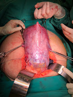
Haematocolpos

What is Haematocolpos: It is a medical condition in which the vagina (colpos) is filled/pooled  with menstrual blood. It is  due to  the blockage of menstrual blood flow. The word hematocolpos stands for  'an accumulation of blood within the vagina'. It is often caused by obstruction to the outflow of menstrual blood flow through the vagina. Haematometra which is the collection of blood in the uterus is sometime related with haematocolpos. The intense pressure & back flow of the obstructed blood leads to formation of Haematometra. In this articles, i will confine our discussion to simple Haematocolpos only.  Below are example causes of haematocolpos. 

a. Imperforate hymen


b. vaginal atresia


c. transverse septum


d. Mullerian Duct Anomalies


Typical Clinical presentation are as below: all these case were manged by me. These cases presented in different scenarios with different physical findings. Tha manage are tailored based on the underlying cause/pathology and was dealt accordingly.

Case 1:

A 12 year old girl, presented to casualty with severe lower abdominal pain. She has this cyclical pain for few months before. She has not attain menarche yet. On examination, the secondary sex characteristics are present. On abdominal examination, there was a 16 week size pelvic mass & its tender on palpation. Perineal examination, there was bluish bulging from the vaginal out let. Pelvic Ultrasound sound showed a large Haematocolpos. The uterus was normal with slight thickened ET & there was some fluid in POD. DX: Imperforate Hymen. Investigation is usually pelvic ultrasound is sufficient. Treatment is usually a simple cruciate incision or elliptical incision of the hymen. Hymen can usually be spaced (hymen spacing surgery). In this case a cruciate incision was made. The staled blood was drained passively. Patient recovered uneventfully.


Case 2:

13 year old girl presented to the casualty with primary amenorrhea & cyclical abdominal pain and difficulty pass urine. On examination, the sexual characteris was normal. The abdomen was distended. Vaginal examination. there were no vaginal opening. There was no perineal bulging. PR there was a gap in between the forchette and the the upper border of the vaginal. The pelvic ultrasound showed a large haematocolpos and some haematometra ( blood collection in the uterus). DX:  Vaginal Atresia


Investigation: pelvic Ultrsaound/ MRI


Treament: Vaginaplasty & Recreation of vagina.
In this case, There was a 2.5 cm of fibrous tissue noted between the vaginal outlet and the lower margin of haematocolpos. The large haematocolpos has helped in stretching the vagina and made the recreation of vaginal easier. Care has to be taken to identify the anterior & Posterior structures. A CBD is useful to assess the anatomy of the urethra. A large bore needle and syringe can be used to find the correct plan between the outlet & haematocolpos. When the correct plane is identified, than, one can use a fine scissors to create a tunnel along the needle to reach the haematocolpos. The upper vaginal margin can be opened & the Haematocolpos can be drained. The upper vaginal margin are than stretched & reattached with the forchette/ or the vaginal outlet. A small fenton operation may be necessary in some cases to facilitate the attachment. The create tunnel/vaginal than be dilated with large hegar dilators to provide a reasonable vaginal diameter.  Patient are also advised to continue to dilated the vagina once the healing process taken place. Dilatation prevents vaginal stenosis.  

Case 3: 
 
!2 plus girl presented with recurrent pelvic pain, aw vomiting & urinary frequency. Examination revealed mass in the lower abdomen with tense swelling. VE finding showed a small vaginal opening & there was a thick septum about 1.5 com from the vaginal outlet. The hymen was seen. A diagnosis of complete transverse septum was made. The septum was excised & the haematocoplos was released without any issues. Patient recovered well. She obtained her  regular periods after the procedure & there were no further heamatocolpos.


Case 4. 

33 yrs, patient who was married for 7 year.  c/o chronic pv discharge & occasional pelvic pain and primary infertility. Has normal periods monthly. No dysmenorrhea. Abdominal examination: flabby abdomen. VE= deep vagina, difficult to see the cervix. moderate bulging at the left vaginal wall.  Ultrasound of pelvis: Large ? Haematometra with Didelphys uterus. The left kidney was absent. Both ovaries seen and appeared normal.
CT Scan: Didelphys uterus with large left vaginal collection (Abscess). The Right kidney normal. Left kidney absent. DX: Ohira syndrome: Delayed & atypical presentation (Left Pyocolpos)


Treatment: EUA, Excision of the septum & Diagnostic Laparoscopy was carried out


Conclusion:
The causes for Hematocolpos are diverse. The above 4 causes are the mostly encountered.  The treat is usually surgical. There are different surgical treatments which needs to be undertaken to cure it. In extreme case like congenital cervical atresia, a complete  hysterectomy may be necessary.

For the women who have an imperforate hymen, a minor surgery is sufficient. An incising for the hymenic membrane or hymenotomy is sufficient to facilitate menstrual flow. In some case with tight outlet, patients may be required to insert dilators into the vagina for a few minutes each day for a few days post the surgery to avoid the incision being closed on its own or to maintain it patency. The surgical correction of a transverse septum and vaginal atresia can be difficult if the surgery is not carefully planned. Postoperative complications, such as vaginal stenosis and re-obstruction can occur, especially when the septum/vagina is thick. In such cases long term dilation with vaginal dilators may be necessary.The thickness and location of the septum is most commonly evaluated by transperineal ultrasound or MRI before attempting its resection. Mullerian duct anomalies can be changing & the surgical treatment planned need to be tailored toward relieve the acute symptoms & long term maintenance for the sexual & reproduction function in such patients. In such cases a preoperative assessment with imaging techniques and multi discipline approach may be necessary to provide effective & long term good outcome. 

Wednesday, 16 March 2022

GENITAL INFECTION IN GYNAECOLOGY 

( MEDICAL STUDENTS LECTURE & NOTES) 2022

The Nature and Role of Physiological Vaginal Discharge.

● Normal for woman to have some degree of vaginal discharge. 

● Normal - white to yellowish (d/t oxidation). 

● Contents: Mucous, desquamated epithelial cells, bacteria and fluid from endometrial.

● There is slight odour but it’s not strong. pH: acidic (4-5). 

● The role : ○ To carry away dead cells and bacteria thus keeps the vagina clean.

                    ○ Acidic - act as defense mechanism against pathogens.

COMMON GENITAL INFECTION IN GYNAECOLOGY:

Vulvovaginal Candidiasis

Causal organism: Candida albicans (gram +ve oval yeast). 

Predisposing factors: Pregnancy (40%), DM, high-dose combined OCP, HIV. 

Signs & symptoms: Vulval itching, thick white curdy discharge (vaginal thrush), dyspareunia, dysuria, vulval oedema, redness, normal vaginal pH. 


Diagnosis: High vaginal swab -> gram stain/wet film examination. 

Treatment: Imidazole oral/pessary (oral contraindicated to pregnant women), nystatin cream/pessary, 

Tricomoniasis 

Causal organism: Trichomonas vaginalis (flagellate protozoa). 

Predisposing factors: Multiple sexual partners, unprotected sex. 

Signs & symptoms: Vulval itching, frothy yellowish green discharge, dysuria, strawberry cervix. Diagnosis: 


Investigation: High vaginal swab, μscopy of vaginal discharge, saline wet mount.


Treatment; Metronidazole


Bacterial Vaginosis 

Bacterial Vaginosis Causal organism: Gardnerella vaginalis, Mycoplasma hominis, Bacteroides spp., Mobilincus spp.

Predisposing factors: Multiple sex partners, douching, lack of good lactobacilli. 

Signs & symptoms: Fishy malodourous discharge, more common during menses. 


Diagnosis: Amsel criteria (≥3 criteria for diagnosing bacterial vaginosis) a) Presence of clue cells (stippled appearance) - μscopic examination. b) Creamy greyish white discharge - naked eye. c) Vaginal pH > 4.5. d) Release of a characteristic fishy odour on addition of alkali. 

Treatment: Metronidazole, clindamycin.

Gonorrhoea 

Causal organism: Neisseria gonorrhoeae (gram -ve diplococcus). 

Predisposing factors: Multiple sex partners, early age of onset of sexual activity. 

Signs & symptoms: Greenish mucopurulent discharge, pelvic tenderness, proctitis, rectal bleeding.

Diagnosis: Vaginal swab -> gram stain/Thayer-Martin agar (blood chocolate agar with antibiotics).

Treatment: Cefixime, ceftriaxone, spectinomycin.


Genitourinary Chlamydia

Causal organism: Chlamydia trachomatis (gram -ve, obligate intracellular parasite).

Predisposing factors: Multiple sex partners, early age of onset of sexual activity.

Signs & symptoms: Mucopurulent discharge, postcoital and intermenstrual bleeding, dysuria. Late stage: Conjunctivitis and pneumonia. 

Diagnosis: Nucleic acid amplification technique, RT-PCR, culture. 


Treatment: Doxycycline, azithromycin, erythromycin, amoxicillin.


 Clerking Format for FEMALE PATIENTS (all, Obstetrics little different)

(INTRO)My Patient name is ………………………..,she is a (age)….. old (Race )……….. lady,  Gravida…….. Para……….   IF PREGNANT GRAVIDA ….,  IF NOT PREGNANT & HAS CHILDREN PARA………. .

She is known case of ……………..( any Medical Problem or issues) eg Diabetse for 10 years and Hypertensive for 3 years. If any MEDICAL PROBLEMS

EG Diagnose as pulm TB , 3 months ago and currently on anti Tb treatment

(CHIEF COMPLAIN)

She presented with history of ………………………………etc…EG.she presented with low grade fever & vomiting for 2 days

(HPI): HISTORY OF PRENSENT ILLNESS

Mrs ………. Eg  case describe c/o in detail, ass factors, aggravating & relieving factors

She was well until last Friday when she develop low grade fever, the fever was of sudden onset, there were no chills or rigor. She denied any body aches, headaches or pain behind her eyes( periorbital pain). The fever settle after one day but on the 2nd day she experience severe vomiting

She vomited between 15-120 times, the vomitus is basically food & water she took, there is no bilous or blood in the vomitus. It was non projectile. There were no obvious aggravating factors. The vomiting slowly improved after taking some medication…….???

On further questioning, she denied any diarrhea, abdominal cramps or pain, she has no history of taking food in any unhygienic places.  Etc etc

Inview of this she seek medical advice from the health  clinic (KKIK……..) from there she was referred to serdang hospital for further management……… She has been in the ward for last 2 days. She was put on intravenous fluid & given some medication. She condition has improve since then. She has been investigated for………… &  what is the treatment & management in the hospital……………

SYSTEMIC REVIEW:…………………………………….No headaches, blurring of vision, no body rashes, etc etc related to your history

MENSTRUAL HISTORY

She attained her menarche at the age of 12 ( …….) age. The cycle has been regular with 28-30 cycles. Her flow is between 5-7 days. There were no menorrhagia (heavy period) or dysmenorrhea (painful periods). Her LMP (last Normal Menstrual Period ) is on the 12. October 2016. She is up to date with her pap smears. Her last pap smear was done in 2016, and she was told it was normal

PAST OBSTETRICS HISTORY

She is para 2, has 2 children age 7 & 2. Both the children were delivered by caesarean section for breech presentation ( baby upside down). The weigh 3 & 3.3 kg. There were no intrapartum & postpartum ( during & after delivery) complications. Both the children are well & alive. She breast fed her children for 6 months….( breast feeding or bottle feeding or mix)

In between pregnancy she took contraception ( OCP, IUCD etc…..)

(PAST MEDICAL HISTORY)

In terms of his past medical hIstory,  He  was diagnosed to have PTb in Sandakan , She was diagnose PTB after she presented with chronic cough & history of LOA, LOW…………, Full assessment was carried out & the diagnosis was confirmed by + sputum for AFB. She was subsequently started with anti-Tb treatment. She has taken the anti TB treatment for last 3 months at the chest clinic/ Health clinic. She is very compliant to the treatment. All her family members were screened & they tested negative.

 SHE IS NOT ALLERGIC TO ANY MEDICATIONS OR FOOD ( Better to put after medical h/o so don’t forget)

 PAST SURGICAL HISTORY

She never had any operations before………

If got operations…….. ? what op & years any complications ( eg……. She had 2 CSection on 2009 & 2017)

 FAMILY HISTORY

She is the youngest of 8 siblings / the third of 4 siblings. His parents are still alive & well. His mother is also diabetic but not hypertensive. His father has no medical illness. All his sibling is well and has no similar illness. There were no one in her family with PTB

 PERSONAL/SOCIAL HISTORY

She is married to EN………………………, she works as a………………., her partner works as……………………….. they live in a flat/ single storey teres / double storey teres/ single or double storey bungalow/condominium I ……………………………kajang/serdang………. The house is well  equipped with adequate basic amenities like water & electricity. Their combine monthly income is RM 2000. They have ……….children. She  and her partner does not smoke cigarette or consume alcohol. He also does not indulge in high risk behaviors’. She take s balance diet with 2-3 serving of meat per week. Recently after contracting with PTB she had loss about 3 kg

 IN SUMMARY, MY PATIENT  Pn/madam…………….., A KNOWN CASE OF DIABETES PTb diagnosed 3 months ago & still on anti-TB treatment presented with low grade fever with severe vomiting

 PROVISIONAL DIAGNOSIS:……………………………………………….

 DIFFERENTIAL DIAGNOSIS:1 …………………………………………

                                                        2………………………………………….

                                                       3. …………………………………………

HOW WOULD YOU MANAGE:………………………………………..

I would like to do some investigation:, l would like to do FBC to see any evidence of infection. I also would like to check his renal function to see if there any evidence of renal impairment. LFT to look for hypoalbuminemia. As per symptoms an ECG & Chest x-ray may provide me with any evidence of pleural effusion or cardiac failure. Others…………………………..

1.       Blood test FBC, RP, LFT …………whey & reasons

2.       Sputum AFB, C&S

3.       Cxray, ecg, echo whey & interpretations

4.       Other test d-dimer, Doppler studies, CT scan, MRI etc………..

5. The options for this patient are A. CONSERVATIVE MANAGEMENT LIKE...

                                                          B. MEDICAL TREATMENT LIKE.....

                                                          C. SURGICAL OPTIONS INCLUDING .........


Wednesday, 21 July 2021

CYSTOSCOPY IN WOMEN

A non invasive procedure to visualize the lower urinary tract system. Mainly credited to Kelly for developing the female cystoscopy.  Hopkin’s introduce the fiberoptic telescopy in 1950’s.

Cystoscopy divided into:

  • Urethroscopy
  • Rigid cystoscopy
  • Flexible cystoscopy

 Intrumentation: 

  • Telescopy ( 0, 30, 70 degrees)
  • Sheathes
  • Bridges
  • Rigid cystoscopy
  • Distension media: Water or Normal saline
  • Other accessory instruments: biopsy forceps, grasping forceps, scissors, stone crusher, Ellipe hydrostatic bottle, electrocautery system. 


Indications: Diagnostic or operative

Diagnostic: Investigative and diagnostic tool for symptoms & signs for urinary problems

  •  Investigation for microscopic or gross haematuria
  •  Infection ( acute or chronic, recurrent)
  •  Inflammation like Interstitial cystitis, radiation cystitis
  •  Overactive bladder symptoms
  •  Voiding dysfunction symptoms
  •  Suspected fistulas
  •  Assessment for staging of cervical cancer  
Operative Procedures: Treat bladder conditions or diseases

  • Periurethral collagen injection for USI/ ISD
  • Intravesicle injections of steroids, botox for intractable DI/ IC
  • Removal of small bladder calculi
  • Removal of foreign bodies like sutures, tape/mesh, polyps & masses
  • Biopsy of abnormal area / tumors

Procedure: https://www.youtube.com/watch?v=pjfXBximSBQ

Cystoscopy may be done using a local anesthetic using lignocaine jelly, under sedation or under general anesthesia in some cases esp patients requiring operative procedure

  • Prior to cystoscopy, empty the bladder
  • Assemble cystoscopy as required flexible or rigid (need to assemble the outer sheath, telescope & the bridge)
  • Test the cystoscopy system in good working condition & white- balance it. 
  • Introduce the cystoscopy with slow water flow to expel any air & to  facilitate the introduction of the tip of scope smoothly through the external meatus & the urethra. inflate the bladder with NS/water up to 200-400mls to get full view of the bladder.
  • Inspect the bladder systematically from the doom & walk through the bladder in clock wise manner.
  • look for the ureteric orifices & trigon area. The distance between the 2 ureteric orifices is about 4 cm
  • inspect the bladder neck & as the scope is withdrawn, inspect the urethra ( usually abloy 4cm length)
  • After the procedure, empty the bladder & apply local anaesthetic gels. 
Normal appearance on cystoscopy


Complications:

  • Injuries to the urethra, bladder wall
  • Bladder perforation
  • Bladder infection 3-5%
  • Bleeding esp after biopsies/ operative procedures
  • Pain

Reporting the findings:

Normal appearance of the bladder; the dome with bubble, bothe the ureteric orifices, trigon area & the bladder neck


Abnormality observed on cystoscopy

                 Rain drop sign as in Interstitial cystitis

                                               Foreign body- mesh /TVT tape, stone

                                                               Bladder perforation:
                               
                                                                      
                                                                          Bladder Polyp


Disclaimers: some pictures & video was uploaded from you-tube & google images
The above article is for teaching purpose
I would like to pay credit to all the contributors for the above pictures & videos
consent for exhibition of the pictures & video has been obtain from the patients

References:

1. https://www.youtube.com/watch?v=q1gDwLaz8oI

Thursday, 24 June 2021

SURGERY FOR FEMALE STRESS URINARY INCONTINANCE

 General considerations

1.   Diversity on choice of surgical procedure. Burch colposuspensions/ fascial slings/ MUS/Injectables
2.  Indication should be significant loss of urine creating social or hygienic problem 
3. Most procedures aim to repositioning of bladder neck and urethra in a supported retropubic position. Others aim to provide increased urethral resistance by improving urethral coaptation.

 Preoperative considerations

1.   History taking, physical examination, laboratory testing and imaging. 

2. Thorough evaluation of all pelvic floor organs should be done preoperatively, In some cases full Urodynaemics studies may be indicated.

3.   Perioperative antibiotic treatment in the operating room.

4. General, regional, or local anaesthesia according to the procedure performed and patient condition

5.  Dorsal lithotomy positioning, prepping and draping the abdomen and vagina in a sterile fashion.

6.    Draining the bladder with a Foley catheter at the beginning of the procedures.

 Operative and postoperative general considerations when performing vaginal surgery

1.  Cystourethroscopy performed at the end of the procedure is recommended to verify that the bladder is intact, that no sutures are traversing the urethra or bladder, and that ureters are patent.

2. The vagina may need to be packed at the end of the procedure to facilitate hemostasis in some cases. Packing can be removed postoperative few hours to the next day.

3. Indwelling catheter or suprapubic catheter is left in the bladder in come cases and as when is needed.

 SUBURETHRAL SLING PROCEDURES (MUS)

  1. Suburethral sling is a strip of material that is tunneled underneath the bladder neck and/or proximal urethra or midurethra and then attached to above structure such as rectus facia or pelvic sidewall to create a posterior support, or hammock effect to the bladder neck and proximal urethra.
  2. Slings are used for all kinds of stress urinary incontinence including anatomical urinary stress incontinence(urethral hypermobility) and can be  completely transvaginally, or with combination of transvaginal and abdominal approach.
  3. Sling materials include:
    1. Autologous sling such as fascia lata or rectus abdominis, harvested at the time of surgery
    2. Homologous material such as cadaveric fascia lata
    3. Synthetic slings: Applied retropubically or transobtoratorly.
  4. Minimally invasive sling procedures have been introduced including the tension free vaginal tape using polypropene tapes.

 Suburethral slings

  • Transvaginal tape (TVT) is becoming popular choice as effective minimally invasive anti-incontinence surgery.
  • Long term results of up to 17 years showed comparable objective and subjective cure rate as to Burch colposuspension.
  • The Tension free vaginal Tape (TVT) is the only sling but that is put at the mid-urethra level. Hence, the name Transvaginal Tape (keeping the TVT abbreviation) was suggested.
  • To reduce such complications, The transobturator approach (TOT). Initial results were comparable with TVT with minimal complications.

 Operative technique of TVT (Tension-free vaginal tape) & Transobturator apparoach (TOT)

  • Positioning and preparation as in preoperative consideration for vaginal surgery.
  • Local, regional, or general anesthesia can be used.
  • Two 5mm long abdominal incisions are made 5cm apart just above the superior rim of the pubic bone (TVT). Small stab incision is made at the ischiopubic angle at the same level as the clitoris (TOT).
  • A 1.5cm long vaginal wall incision is made over the midurethra, 1cm proximal to the external urethral meatus.
  • Bilateral paraurethral dissection of vaginal wall is performed.
  • A prolene tape attached on both ends to trocars/needles and covered by a plastic sheath is used. One of the trocars/needles is introduced and advanced through the vaginal incision, the urogenital diaphragm, and the retropubic space in close contact to the posterior aspect of the pubic bone until its tip is brought out to the abdominal incision. TOT has similar approach but the needles are introduced either through inside out as in TVTo or from out side in as other TOT’s.
  • Using the trocar/ needles at the other end of the sling, this step is then repeated, on the contralateral side.
  • Cystoscopy is performed to rule out bladder or urethral damage.
  • Once bladder and urethral integrity have been verified, the trocars/needles on both sides removed and hence the tape is pulled all the way through the abdominal incision. The tape tension is further adjusted under the urethra.
  • The plastic sheath covering of the tape is removed. The friction between the tape and the tissue canal created by the trocars serves to hold the sling in place with no need for additional suture fixation of the sling.
  • Abdominal and vaginal incisions are closed.   

 Complications:

  • Bladder perforation ( 5% with TVT, almost nil with TOT)
  • Bowel Injuries ( 0.7/1000)
  • vascular injuries ( 0.7/1000)
  • Bleeding
  • Voiding dysfunction (2.8% or 23/1000- 79/1000)
  • Overactive bladder.
  • Tape erosion/exposure ( <1%)


References

1. Transobturator tape for Stress Incontinence: North Queensland Experience NAidu A, Lim YN, Barry C et al . Aust N Z J Obstet Gynaecol. 2005 Oct;45(5):446-9

2. Does the MORNAC Transobturator sling cause post-operative voiding dysfunction? A prospective study. Barry C, Naidu A, Lim YN et al. Int Urogynecol J Pelvic Floor Dysfunct. 2006 Jan;17(1):30-4

3. Transobturator sub-urethral suspension, an approach worth changing to?.Malaysian Journal of O & G: 2005 ( Suppl)

4.  Transobturator Tape(TOT) procedure: The Ipoh Experience JUMMEC 2011;14(1)

http://jummec.um.edu.my/filebank/published_article/2967/JuMMEC%202011%2014(1)%2010-20.pdf

 5. Seventeen years' follw-up of the tension-free vaginal tape procedure for stress incontinence. Nilsson CG et al . Int Urogynaecol J 24(8)


Tuesday, 22 June 2021

MISSION ULAAN BAATOR, MONGOLIA

I had an opportunity to provide some training for young doctors & specialist in Ulaan Baator on OASIS & Urogynae update. A team of 4 urogynaecologist ( Me, Dr Ng Poh Yin, Dr Tan GI & Dr Ida Lily Waty) manage to organise & execute this valuable training.  It took us near full one day to reach Ulaan Baator, as there is no direct flights from KL. The Trip was co-organise by MUGS & Mongolian Urogynae society and Mongolian O & G society. During this trip we had lectures on OASIS & wet lab / hands on sessions with the mongolian trainees & specialist. The day 2 was mainly an update in Urogynaecology. The training was well accepted. We had nearly 40 doctors from different parts of Mongolia participated in the training.

Following the training course, we also had life time opportunity to explore deep part of Mongolia. It was vast & unexplored territory. 


Mongolia is a country sandwiched by Russia in the north & china in the south-east. It has vast steppe land. The total population is about 4 million & more than half of them live in Ulaan Baator (Capital of Mongolia). Mongolia was at one time a great country.  Genghis Khan is a great Mongol emperor who had conquered large part of Central Asia and China in  13 & 14th century.


          The Ulaan Baator City is a modern City. The Main hospital is well equipped and up to date.


                     The country has beautiful terrin. The people are super friendly and welcoming. 


Overall it was a great teaching & exploration trip for all of us